2012 AIPMT-MAINS Structural Organisation in AnimalsAnimal Tissues Medium Question 2 of 2 in Animal Tissues

The four sketches (A, B, C and D) given below represent four different types of animal tissues. Which one of…

The four sketches (A, B, C and D) given below represent four different types of animal tissues. Which one of these is correctly identified in the options given, along with its correct location and function?
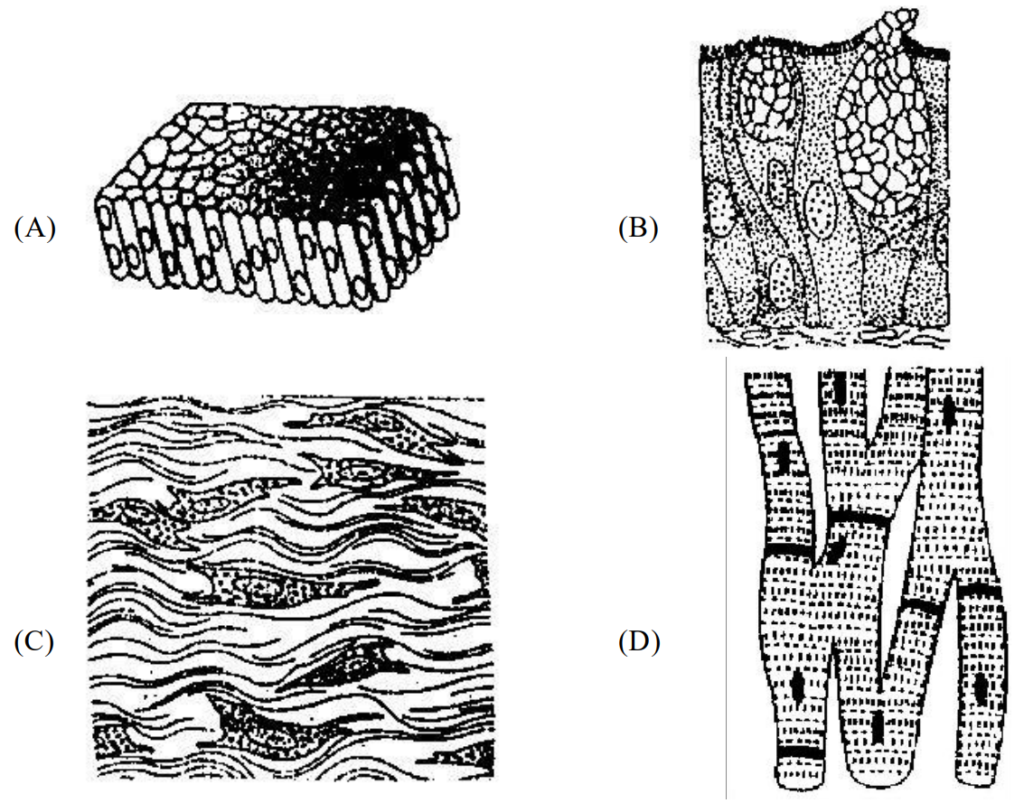
A (A) Columnar epithelium – Nephron – Secretion and absorption
B (B) Glandular epithelium – Intestine – Secretion
C (C) Collagen fibres – Cartilage – Attach skeletal muscles to bones
D (D) Smooth muscle tissue – Heart – Heart contraction

The answer is B.

Solution

Sketch (B) shows glandular epithelium (columnar cells specialised for secretion, including unicellular goblet cells); it occurs in the lining of the intestine and its function is secretion. This row is fully correct.
Sketch (A): columnar epithelium lines the stomach and intestine; the tubules of the nephron are lined by cuboidal epithelium, so the location is wrong.
Sketch (C): the structure that attaches skeletal muscles to bones is a tendon (dense regular connective tissue), not cartilage, so the row is wrong.
Sketch (D): smooth muscle is found in the walls of blood vessels, stomach and intestine; the heart is made of cardiac muscle, so the row is wrong.
Hence the correct identification is (B) glandular epithelium, found in the intestine, with the function of secretion.

Concept Epithelial tissue

Animal Tissues in past papers

2 questions from this chapter have appeared across 2 exam years.