Looking for classes? Ksquare Career Institute, Bengaluru →

Biology
Neural Control and Coordination
61 questions from 34 papers, each with its answer.
- 61 questions
- 34 papers
- 1.73% of Biology
Functions of Pons, Hypothalamus, Medulla and Cerebellum
-
Match List I with List II regarding parts of the brain and their functions.
Pick the matching item from Column II for each row.
Column I
- A. Pons
- B. Hypothalamus
- C. Medulla
- D. Cerebellum
Column II
- I. Provides additional space for neurons, regulates posture and balance.
- II. Controls respiration and gastric secretions.
- III. Connects different regions of the brain.
- IV. Neuro secretory cells
Correct answer: A → III, B → IV, C → II, D → I
Pons – consists of fibre tracts that interconnect different regions of the brain Hypothalamus – contains several groups of neurosecretory cells that secrete hypothalamic hormones Medulla – contains centres that control respiration and gastric secretions Cerebellum – convoluted surface provides additional space for neurons; regulates posture and balance Correct match: A-III, B-IV, C-II, D-I.
Generation and Conduction of Nerve Impulse – Synapse
-
Select the incorrect statement regarding synapses:
Sense Organs – The Ear
-
Match the following columns and select the correct option.
Pick the matching item from Column II for each row.
Column I
- A. Organ of Corti
- B. Cochlea
- C. Eustachian tube
- D. Stapes
Column II
- I. Connects middle ear and pharynx
- II. Coiled part of the labyrinth
- III. Attached to the oval window
- IV. Located on the basilar membrane
Correct answer: A → IV, B → II, C → I, D → III
Sense Organs – The Human Eye
-
Which of the following statements is correct?
Neuron structures and locations
-
Match List-I with List-II and choose the correct answer from the options given below:
Pick the matching item from Column II for each row.
Column I
- A. Schwann cells
- B. Synaptic knob
- C. Bipolar neurons
- D. Multipolar neurons
Column II
- I. Neurotransmitter
- II. Cerebral cortex
- III. Myelin sheath
- IV. Retina
Correct answer: A → III, B → I, C → IV, D → II
Schwann cells form the myelin sheath around axons (A-III); Synaptic knob releases neurotransmitter at the synapse (B-I); Bipolar neurons are found in the retina (C-IV); Multipolar neurons are found in the cerebral cortex (D-II).
Structures and functions of the eye
-
Match the following columns and select the correct option:
Pick the matching item from Column II for each row.
Column I
- a. Rods and Cones
- b. Blind Spot
- c. Fovea
- d. Iris
Column II
- i. Absence of photoreceptor cells
- ii. Cones are densely packed
- iii. Photoreceptor cells
- iv. Visible coloured portion of the eye
Correct answer: a → iii, b → i, c → ii, d → iv
Rods and Cones are photoreceptor cells (a-iii); Blind Spot shows absence of photoreceptor cells (b-i); Fovea has densely packed cones (c-ii); Iris is the visible coloured portion of the eye (d-iv).
Olfactory receptors and structure of the eye
-
Given below are two statements:- Statement I: The nose contains mucus – coated receptors which are specialised for receiving the sense of smell and are called olfactory receptors. Statement II: Wall of the eye ball has three layers. The external layer is called choroid (dense connective tissue), middle layer is sclera (thin pigmented layer) and internal layer is retina (ganglion cells, bipolar cells and photoreceptor cells). In the light of the above statements, choose the correct answer from the options given below:Statement I correctly describes olfactory receptors in the nose. Statement II incorrectly swaps the names/descriptions of the eyeball's layers: the external layer is actually the sclera (dense connective tissue), and the middle layer is the choroid (thin, pigmented, vascular layer) — the layers given are reversed, so Statement II is false.
Structure of the brainstem
-
Brainstem of human brain consists of:The brainstem consists of the midbrain, pons, and medulla oblongata.
Types of neurons and nerve fibres
-
Match List - I with List - II. Choose the correct answer.
Pick the matching item from Column II for each row.
Column I
- A. Multipolar neuron
- B. Bipolar neuron
- C. Myelinated nerve fibre
- D. Unmyelinated nerve fibre
Column II
- I. Somatic neural system
- II. Cerebral cortex
- III. Retina of Eye
- IV. Spinal nerves
Correct answer: A → II, B → III, C → IV, D → I
Multipolar neurons (one axon, two or more dendrites) are found in the cerebral cortex; bipolar neurons are found in the retina of the eye; myelinated nerve fibres are found in spinal and cranial nerves; unmyelinated nerve fibres are found in the autonomous and somatic neural systems.
Nerve impulse, reflex action and photoreceptors
-
Which of the following statements is not correct?
Human brain – parts and functions
-
Which of the following structures or regions is incorrectly paired with its functions?
The limbic system regulates emotion, not movement.
The limbic system is the emotional brain. It controls emotions in our body but not movements. So the limbic system is incorrectly paired.
Sense organs – photoreceptors and visual pigments
-
Good vision depends on adequate intake of carotene rich food. Select the best option from the following statements (a) Vitamin A derivatives are formed from carotene (b) The photopigments are embedded in the membrane discs of the inner segment (c) Retinal is a derivative of vitamin A (d) Retinal is a light absorbing part of all the visual photopigments
Statements (a), (c) and (d) are correct; pigments lie in the outer segment.
Carotene is the source of retinal, which is involved in the formation of rhodopsin in rod cells. Retinal, a derivative of vitamin A, is the light-absorbing part of all visual photopigments. Photopigments are in the membrane discs of the outer segment, so (b) is incorrect.
Human eye
-
Parts A, B, C and D of the human eye are shown in the diagram. Select the option which gives correct identification along with its functions/characteristics:
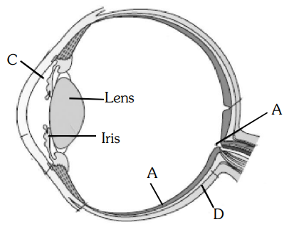 A is the retina, the inner layer that contains the photoreceptor cells, rods and cones.
The blind spot has no photoreceptors, and the anterior part of the choroid forms the ciliary body.
Spinal cord
-
Destruction of the anterior horn cells of the spinal cord would result in loss of:
Synapse
-
A diagram showing axon terminal and synapse is given. Identify correctly at least two of A-D.
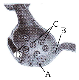 A is the receptor on the post-synaptic membrane and C is the synaptic vesicle containing neurotransmitter.
B is the synaptic cleft and D is $Ca^{++}$.
Generation and conduction of nerve impulse
-
During the transmission of nerve impulse through a nerve fibre, the potential on the inner side of the plasma membrane has which type of electric change?In the resting (polarised) state the inner side of the plasma membrane of the nerve fibre is negatively charged with respect to the outside; the resting potential is about -70 mV.
When a stimulus is applied, the membrane at that site becomes freely permeable to $Na^+$, and $Na^+$ ions rush in.
The polarity is reversed: the inner side becomes positive (about +30 mV). This is depolarisation, and the potential difference is the action potential, which lasts about 1 millisecond.
Immediately afterwards the permeability to $K^+$ increases and $K^+$ ions diffuse out, so the inner side becomes negative again. This is repolarisation; the sodium-potassium pump then restores the original ionic distribution.
The same sequence is repeated at each successive point as the impulse travels along the fibre.
So the potential on the inner side is first negative, then positive and again back to negative. -
Depolarization of axolemma during nerve conduction takes place because of :At rest, the inside of the axon is negative (about –70 mV).
When stimulated, voltage-gated $Na^+$ channels open and $Na^+$ ions rush into the axon, so the inside becomes positive (depolarisation, about +30 mV).
Then the $K^+$ channels open and $K^+$ moves out, restoring the negative potential (repolarisation).
So, as per the answer key, depolarisation involves $Na^+$ moving inside, with $K^+$ moving outside afterwards.
Human brain and sense organs
-
Select the answer with correct matching of the structure, its location and function.The hypothalamus lies at the base of the thalamus, which is part of the forebrain; it contains centres that control body temperature and the urge for eating and drinking. This row is fully correct.
The Eustachian tube does equalise the pressure on either side of the ear drum, but it connects the middle ear cavity with the pharynx; it is not part of the internal ear.
The cerebellum is part of the hindbrain and maintains balance and posture; respiration and gastric secretions are controlled by the medulla.
The blind spot is the point where the optic nerve leaves the eye and the retinal blood vessels enter it; photoreceptor cells are not present in that region.
Hence the correct match is hypothalamus – forebrain – controls body temperature, urge for eating and drinking.
Neurotransmitters
-
Alzheimer disease in humans is associated with the deficiency ofAlzheimer's disease is a degenerative disorder of the brain marked by progressive loss of memory and of intellectual functions.
It is associated with the degeneration of the cholinergic neurons of the brain and a marked fall in the level of the neurotransmitter acetylcholine.
Deficiency of dopamine is associated with Parkinson's disease.
Hence Alzheimer's disease is associated with the deficiency of acetylcholine.
Structure of the Ear
-
Given below is a diagrammatic cross section of a single loop of human cochlea:
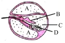
Which one of the following options correctly represents the names of three different parts?In a cross section, each loop of the cochlea shows three canals.
The upper canal is the scala vestibuli and the lower canal is the scala tympani; both are filled with perilymph. In the figure both of these outer chambers are labelled A, so A is perilymph.
The middle canal is the scala media (cochlear duct), which is filled with endolymph. It is separated from the scala vestibuli by Reissner's membrane and from the scala tympani by the basilar membrane. This is the part labelled C, so C is endolymph.
The organ of Corti is located on the basilar membrane and contains the sensory hair cells that act as auditory receptors.
Above the rows of hair cells is a thin elastic membrane called the tectorial membrane, labelled B.
The hair cells are sensory cells, not secretory cells, and the fluid in the outer canals is perilymph, not endolymph or serum.
So the correct labelling is A: Perilymph, B: Tectorial membrane, C: Endolymph.
Sense Organs: Olfactory Receptors
-
Bowman's glands are located in theBowman's glands (olfactory glands) are located in the olfactory epithelium (olfactory mucosa) lining the upper part of the nasal cavity.
They secrete mucus that keeps the olfactory epithelium moist.
Odorous substances dissolve in this mucus and are then detected by the olfactory receptor cells; the secretion also washes the surface clean for fresh stimuli.
They should not be confused with Bowman's capsule, the cup-shaped structure at the proximal end of the uriniferous tubule (nephron) that encloses the glomerulus.
They are not found in the anterior pituitary or in the reproductive system of the cockroach.
So Bowman's glands are located in the olfactory epithelium of our nose. -
Bowman's glands are found in -Bowman's glands (olfactory glands) are found in the olfactory epithelium lining the upper part of the nasal cavity.
They secrete mucus that keeps the olfactory epithelium moist.
Odorous substances dissolve in this mucus before they stimulate the olfactory receptor cells; the secretion also washes the surface clear for new odours.
They should not be confused with Bowman's capsule, the cup-like part of the nephron that surrounds the glomerulus.
The external auditory canal has ceruminous (wax) glands, not Bowman's glands.
So Bowman's glands are found in the olfactory epithelium.
Autonomic Nervous System
-
One of the examples of the action of the autonomous nervous system is -The autonomic nervous system controls the involuntary activities of the internal organs (viscera) and smooth muscles.
Peristalsis of the intestines is a wave of contraction of the smooth muscles of the gut wall; it is regulated by the autonomic nervous system (the parasympathetic system increases it and the sympathetic system decreases it).
The knee-jerk response is a spinal reflex involving skeletal muscle, controlled by the somatic nervous system.
Swallowing begins as a voluntary action of skeletal muscles.
The pupillary reflex also involves autonomic fibres, but the answer accepted by the key is peristalsis.
So the example of the action of the autonomic nervous system is the peristalsis of the intestines.
Neurotransmitters and Brain Disorders
-
Parkinson's disease (characterized by tremors and progressive rigidity of limbs) is caused by degeneration of brain neurons that are involved in movement control and make use of neurotransmitter -Parkinson's disease is a degenerative disorder of the brain, marked by tremors, rigidity of the limbs and slowness of movement.
It is caused by the degeneration of the dopamine-producing neurons of the substantia nigra in the midbrain.
These neurons send fibres to the basal ganglia (corpus striatum), which help to control and smooth out body movements.
With the loss of dopamine, this control is disturbed and the characteristic symptoms appear.
The disease is treated with L-dopa, a precursor that is converted into dopamine in the brain.
So the neurotransmitter involved is dopamine.
Resting Membrane Potential
-
In the resting state of the neural membrane, diffusion due to concentration gradients, if allowed, would drive :-At rest, the sodium-potassium pump maintains a high concentration of $Na^+$ outside the axon and a high concentration of $K^+$ inside.
Diffusion always moves an ion from higher to lower concentration.
So $Na^+$, being more concentrated outside, would diffuse into the cell.
$K^+$, being more concentrated inside, would diffuse out of the cell (not into it).
Among the given choices, the correct one is that diffusion would drive $Na^+$ into the cell. (At rest the membrane is far less permeable to $Na^+$, which is why this does not happen until an action potential opens sodium channels.)
Peripheral Nervous System: Cranial Nerves
-
Injury to vagus nerve in humans is not likely to affect -The vagus nerve (tenth cranial nerve) is a mixed nerve and the main parasympathetic nerve of the body.
It supplies the heart (slows the heart rate), the larynx and pharynx, the lungs, and the gastrointestinal tract (it increases peristalsis and secretion of digestive juices, including pancreatic secretion).
So an injury to the vagus nerve can affect cardiac movements, gastrointestinal movements and pancreatic secretion.
Movements of the tongue are controlled by the hypoglossal nerve (twelfth cranial nerve), not by the vagus.
So injury to the vagus nerve is not likely to affect tongue movements.
That is every question in Neural Control and Coordination for this filter.
Show all 61 questions Next: Chemical Coordination and Integration
Want a score?
Answers here are not saved. Practise as a test to be marked and see your progress.
Practise as a test