Looking for classes? Ksquare Career Institute, Bengaluru →

Biology
Body Fluids and Circulation
83 questions from 38 papers, each with its answer.
- 83 questions
- 38 papers
- 2.35% of Biology
Cardiac Conduction System
-
Following are the stages of pathway for conduction of an action potential through the heart: A. AV bundle B. Purkinje fibres C. AV node D. Bundle branches E. SA node Choose the correct sequence of pathway from the options given below:The SA node generates the action potential, which stimulates both atria to contract. It is conducted to the AV node, then through the AV bundle (bundle of His), which divides into the right and left bundle branches, and finally through the Purkinje fibres into the ventricular musculature. Pathway: SA node → AV node → AV bundle → Bundle branches → Purkinje fibres, i.e. E-C-A-D-B.
Electrocardiogram (ECG) Waves
-
Match List-I with List-II regarding ECG waves and the cardiac event they represent.
Pick the matching item from Column II for each row.
Column I
- A. P wave
- B. QRS complex
- C. T wave
- D. T-P gap
Column II
- I. Heart muscles are electrically silent.
- II. Depolarisation of ventricles.
- III. Depolarisation of atria.
- IV. Repolarisation of ventricles.
Correct answer: A → III, B → II, C → IV, D → I
P wave – depolarisation of atria QRS complex – depolarisation of ventricles T wave – repolarisation of ventricles T-P gap – heart muscles are electrically silent Correct match: A-III, B-II, C-IV, D-I.
Electrocardiograph (ECG)
-
Match List-I with List-II.
Pick the matching item from Column II for each row.
Column I
- A. P-wave
- B. Q-wave
- C. QRS complex
- D. T-wave
Column II
- I. Beginning of systole
- II. Repolarisation of ventricles
- III. Depolarisation of atria
- IV. Depolarisation of ventricles
Correct answer: A → III, B → I, C → IV, D → II
The P-wave is atrial depolarisation, QRS is ventricular depolarisation and the T-wave is ventricular repolarisation.
In an ECG record, three recognisable waves (P-wave, QRS complex and T-wave) appear with each heartbeat. P-wave – represents atrial depolarisation. QRS complex – represents ventricular depolarisation. T-wave – represents ventricular repolarisation. Q-wave – represents initial phase of ventricular depolarisation i.e., beginning of systole. -
Match Column-I with Column-II.
Pick the matching item from Column II for each row.
Column I
- A. P-wave
- B. QRS complex
- C. T-wave
- D. Reduction in the size of T-wave
Column II
- I. Depolarisation of ventricles
- II. Repolarisation of ventricles
- III. Coronary ischemia
- IV. Depolarisation of atria
- V. Repolarisation of atria
Correct answer: A → IV, B → I, C → II, D → III
Blood – Formed Elements (WBCs)
-
Which of the following statements are correct? A. Basophils are most abundant cell of the total WBCs B. Basophils secrete histamine, serotonin and heparin C. Basophils are involved in inflammatory response D. Basophils have kidney shaped nucleus E. Basophil are agranulocyte Choose the correct answer from the options given below:
Basophils are the least abundant WBCs and are granulocytes that secrete histamine, serotonin and heparin.
Neutrophils are the most abundant cells (60–65%) of the total WBCs and basophils are the least (0.5–1%). Basophils have S-shaped nucleus. Neutrophils, eosinophils and basophils are different types of granulocytes. -
Match the following columns and select the correct option.
Pick the matching item from Column II for each row.
Column I
- A. Eosinophils
- B. Basophils
- C. Neutrophils
- D. Lymphocytes
Column II
- I. Immune response
- II. Phagocytosis
- III. Release histaminase, destructive enzymes
- IV. Release granules containing histamine
Correct answer: A → III, B → IV, C → II, D → I
Blood – Coagulation; Spleen
-
Given below are two statements: Statement I: The coagulum is formed of network of threads called thrombins Statement II: Spleen is the graveyard of erythrocytes In the light of the above statements, choose the most appropriate answer from the options given below.
Cardiac Cycle
-
Which one of the following statements is correct?
-
In humans, blood passes from the post caval to the diastolic right atrium of heart due to:Blood always flows from a region of higher pressure to a region of lower pressure.
During atrial diastole the right atrium is relaxed, so the pressure inside it falls to nearly zero.
The pressure of the blood in the great veins (the post caval or inferior vena cava) is low, but it is still higher than the pressure in the relaxed atrium.
Because of this pressure difference between the post caval and the atrium, blood flows passively from the vein into the right atrium.
The sino-auricular node initiates the contraction (systole) of the atria; it does not bring blood into the atrium.
There are no valves at the openings of the venae cavae that need to be pushed open, and the filling is due to the pressure gradient.
So blood passes into the diastolic right atrium due to the pressure difference between the post caval and the atrium.
Cardiac Cycle – Cardiac Output
-
Which would be the heart rate of a person if the cardiac output is 5 L, blood volume in the ventricles at the end of diastole is 100 mL and at the end of ventricular systole is 50 mL?
WBC differential count
-
The WBC count of a person's blood sample is 8000/cu.mm. How many eosinophils and lymphocytes would be in the same blood sample approximately?
ABO and Rh blood group systems
-
A person with blood group $ARh^-$ can receive the blood transfusion from which of the following types? A. $BRh^-$ B. $ABRh^-$ C. $ORh^-$ D. $ARh^-$ E. $ARh^+$. Choose the correct answer from the options given below:A recipient with $Rh^-$ blood cannot receive $Rh^+$ blood (would trigger an immune response against the Rh antigen). Also, as an A blood group individual has anti-B antibodies in plasma, they cannot receive blood containing the B antigen (B or AB groups). Hence the donor can only be $O\,Rh^-$ (universal donor, C) or $A\,Rh^-$ (D) — so the answer is C and D only.
Open vs closed circulatory systems
-
Open Circulatory system is present in:Anopheles (an arthropod), Limax (a mollusc) and Limulus (an arthropod) all possess an open circulatory system, in which blood does not always flow through vessels but bathes tissues directly.
Cardiac cycle and heart sounds
-
'Lub' sound of Heart is caused by the ________.The first heart sound ('lub') is produced by the closure of the atrioventricular (tricuspid and bicuspid/mitral) valves at the start of ventricular systole.
Neural regulation of cardiac output
-
Which of the following is associated with decrease in cardiac output?Parasympathetic neural signals (via the vagus nerve) decrease the heart rate and cardiac output, opposing the effect of sympathetic stimulation and adrenal medullary hormones, which increase cardiac output.
ECG waveforms and their significance
-
Match List-I (ECG) with List-II (Electrical activity of heart).
Pick the matching item from Column II for each row.
Column I
- A. P-wave
- B. QRS complex
- C. T wave
- D. End of T wave
Column II
- I. Depolarisation of ventricles
- II. End of systole
- III. Depolarisation of atria
- IV. Repolarisation of ventricles
Correct answer: A → III, B → I, C → IV, D → II
P-wave represents depolarisation of the atria (A-III); QRS complex represents depolarisation of the ventricles (B-I); T wave represents repolarisation of the ventricles (C-IV); the end of the T wave marks the end of systole (D-II).
Differential white blood cell count
-
Match List-I with List-II.
Pick the matching item from Column II for each row.
Column I
- A. Eosinophils
- B. Lymphocytes
- C. Neutrophils
- D. Monocytes
Column II
- I. 6-8%
- II. 2-3%
- III. 20-25%
- IV. 60-65%
Correct answer: A → II, B → III, C → IV, D → I
Eosinophils make up 2-3% of white blood cells (A-II); Lymphocytes make up 20-25% (B-III); Neutrophils make up 60-65% (C-IV); Monocytes make up 6-8% (D-I).
Formed elements of blood
-
Arrange the following formed elements in the decreasing order of their abundance in blood in humans: (a) Platelets (b) Neutrophils (c) Erythrocytes (d) Eosinophils (e) Monocytes Choose the most appropriate answer from the options given below:Erythrocytes: 5-5.5 million/mm$^3$; platelets: 1.5-3.5 lakh/mm$^3$; WBCs: 6000-8000/mm$^3$, of which neutrophils 60-65%, monocytes 6-8%, eosinophils 2-3%. Order: erythrocytes > platelets > neutrophils > monocytes > eosinophils.
-
There is no DNA inMature mammalian RBCs lose their nucleus during development, so they have no nuclear DNA; they also lack mitochondria, so they have no mitochondrial DNA either.
Hair root cells are living, nucleated cells and contain DNA.
An enucleated ovum has lost its nucleus but still has mitochondria in its cytoplasm, which contain mitochondrial DNA.
A mature spermatozoan has a haploid nucleus in its head and mitochondria in its middle piece.
Hence there is no DNA in mature RBCs.
Nodal tissue - AV node
-
A specialised nodal tissue embedded in the lower corner of the right atrium, close to Atrio-ventricular septum, delays the spreading of impulses to heart apex for about 0.1 sec. The delay allows
Coagulation of blood
-
You are required to draw blood from a patient and to keep it in a test tube for analysis of blood corpuscles and plasma. You are also provided with the following four types of test tubes. Which of them will you not use for the purpose?To study blood corpuscles and plasma, the blood must be kept from clotting.
Calcium ions ($Ca^{2+}$) are essential for blood coagulation (they are needed for the conversion of prothrombin into thrombin and in other steps of the clotting cascade).
A test tube containing calcium bicarbonate would supply extra $Ca^{2+}$ ions and promote clotting, so it must not be used.
Sodium oxalate (and citrate) removes $Ca^{2+}$ by forming insoluble calcium oxalate, so it prevents clotting.
Heparin is a natural anticoagulant that prevents the conversion of prothrombin into thrombin.
Chilling slows down the enzymatic reactions of clotting, so a chilled tube also delays coagulation.
So the test tube containing calcium bicarbonate is the one you will not use.
Formed elements of blood – erythrocytes
-
Adult human RBCs are enucleate. Which of the following statement(s) is/are most appropriate explanation for this feature? (a) They do not need to reproduce (b) They are somatic cells (c) They do not metabolize (d) All their internal space is available for oxygen transport
Loss of nucleus maximises space for haemoglobin.
In human RBCs, the nucleus degenerates during maturation, which provides more space for the oxygen-carrying pigment (haemoglobin). RBCs lack most cell organelles, including mitochondria, so they respire anaerobically (they do metabolise).
Myogenic heart
-
Frog's heart when taken out of the body continues to beat for some time. Select the best option from the following statements (a) Frog is a poikilotherm (b) Frog does not have any coronary circulation (c) Heart is "myogenic" in nature (d) Heart is autoexcitable
A myogenic, autoexcitable heart beats without nervous input.
The frog (like other vertebrates) has a myogenic heart with a self-contractile system, i.e. it is autoexcitable; because of this it keeps working outside the body for some time.
Blood vessels – blood pressure
-
Blood pressure in the pulmonary artery is
Arterial pressure exceeds venous pressure.
Blood pressure in the pulmonary artery is more than in the pulmonary vein; hence the wall of the pulmonary artery is thick and elastic, while the pulmonary vein walls are thin.
Formation of RBCs
-
Erythropoiesis starts inErythropoiesis begins in the embryonic stage — first in the yolk sac, then in the liver and spleen, and later in red bone marrow.
In adults it takes place in the red bone marrow, stimulated by erythropoietin released by the kidneys.
Double circulation
-
Figure shows schematic plan of blood circulation in humans with labels A to D. Identify the label and give its function/s.
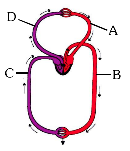 C is the vena cava, which brings deoxygenated blood from the body parts to the right auricle; its $PCO_2$ is 45 mm Hg.
The pulmonary vein carries oxygenated blood and the pulmonary artery carries deoxygenated blood. -
Systemic heart refers to : -In birds and mammals (higher vertebrates), the heart is four-chambered and the left and right halves are completely separated, giving double circulation.
The right side (right atrium and ventricle) receives deoxygenated blood and pumps it to the lungs; it is called the pulmonary heart.
The left side (left atrium/auricle and left ventricle) receives oxygenated blood from the lungs and pumps it to all parts of the body through the aorta (systemic circulation). So it is called the systemic heart.
In fishes, the whole heart pumps only deoxygenated blood (venous heart).
So the systemic heart refers to the left auricle and left ventricle in higher vertebrates.
Electrocardiograph
-
Given below is the ECG of a normal human. Which one of its components is correctly interpreted below?
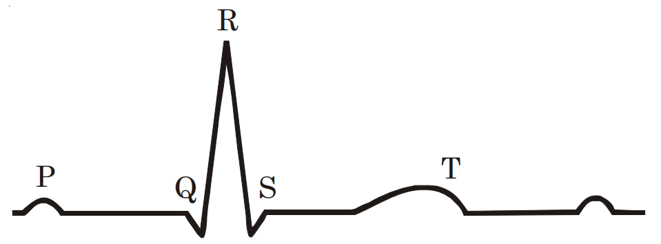 The P-wave represents the electrical excitation (depolarisation) of the atria, which leads to the contraction of both atria, not of the left atrium only.
The QRS complex represents the depolarisation of the ventricles, which initiates ventricular contraction (systole).
Each ventricular contraction pushes blood into the arteries and produces one pulse; so by counting the QRS complexes in a given time one can find the heart beat (pulse) rate. Thus one QRS complex corresponds to one complete pulse.
The T-wave represents the return of the ventricles from the excited to the normal state (repolarisation); its end marks the end of systole, not the start of contraction.
The ECG records electrical activity; its peaks do not measure systolic and diastolic blood pressures.
Hence the correct interpretation is complex QRS – one complete pulse.
Blood vessels
-
In which point is pulmonary artery different from pulmonary vein :Arteries carry blood away from the heart under high pressure, so they have thick, muscular and elastic walls and a narrow lumen.
Veins carry blood towards the heart under low pressure, so they have thin walls, a wide lumen and valves.
The pulmonary artery (carrying deoxygenated blood to the lungs) is an artery, so its wall is thicker than that of the pulmonary vein.
Both have an endothelium lining.
So the pulmonary artery differs in that its wall is thick.
Human heart
-
If due to some injury the chordae tendinae of the tricuspid valve of the human heart is partially non-functional, what will be the immediate effect?The tricuspid valve guards the opening between the right atrium and the right ventricle.
The chordae tendinae are tendinous cords that attach the flaps of the valve to the papillary muscles of the ventricle; they keep the flaps from turning back into the atrium when the ventricle contracts.
If they are partly non-functional, the valve will not close properly during ventricular systole and some blood will leak back into the right atrium.
Less blood will then be pumped from the right ventricle into the pulmonary artery.
The aorta and the left atrium are on the left side of the heart and are not directly affected, and the pacemaker (SA node) is independent of the valve.
Hence the flow of blood into the pulmonary artery will be reduced.
Erythrocytes
-
G-6-P dehydrogenase deficiency is associated with haemolysis of -Glucose-6-phosphate dehydrogenase (G6PD) is the first enzyme of the pentose phosphate pathway, which produces NADPH.
In red blood cells this pathway is the only source of NADPH, which keeps glutathione in its reduced form; reduced glutathione protects the cells against damage by oxidising agents.
In G6PD deficiency (an X-linked recessive disorder) the RBCs cannot protect themselves from oxidative stress.
On exposure to certain drugs (such as primaquine and sulphonamides), infections or fava beans, the haemoglobin and membranes of the RBCs are damaged and the cells burst.
This haemolysis of red blood cells causes haemolytic anaemia.
So G6PD deficiency is associated with the haemolysis of RBCs.
Erythrocytes and Anaemia
-
Examination of blood of a person suspected of having anemia, shows large, immature, nucleated erythrocytes without haemoglobin. Supplementing his diet with which of the following is likely to alleviate his symptoms ?Large, immature, nucleated red blood cells in the blood are called megaloblasts, and the condition is megaloblastic (macrocytic) anaemia.
The maturation of red blood cells in the bone marrow requires folic acid and vitamin $B_{12}$ (cobalamine), because both are needed for the synthesis of DNA.
When they are deficient, DNA synthesis and cell division are slowed while the cells keep growing, so large, immature, nucleated cells are released into the blood.
Deficiency of vitamin $B_{12}$ in particular causes pernicious anaemia.
So supplementing the diet with folic acid and cobalamine is likely to relieve the symptoms.
Iron deficiency causes a different anaemia, in which the red cells are small and pale (microcytic, hypochromic).
Thiamine ($B_1$) deficiency causes beri-beri and riboflavin ($B_2$) deficiency causes cheilosis.
Conducting System of Heart
-
Bundle of His is a network of : -The conducting system of the heart (SA node, AV node, bundle of His and Purkinje fibres) is made of specialised (modified) cardiac muscle fibres, not nerve fibres. The heart is myogenic.
The bundle of His (AV bundle) starts from the AV node, passes through the interventricular septum, and divides into right and left bundle branches, which give rise to the Purkinje fibres spreading throughout the ventricular walls.
So it carries the impulse only to the ventricles; the atria receive the impulse directly from the SA node through the atrial muscle.
So the bundle of His is a network of muscle fibres found only in the ventricle wall.
That is every question in Body Fluids and Circulation for this filter.
Show all 83 questions Next: Excretory Products and their Elimination
Want a score?
Answers here are not saved. Practise as a test to be marked and see your progress.
Practise as a test